Table of Contents
Bullwinkle Disease in Deer
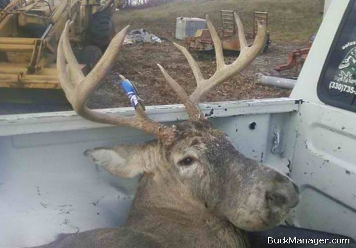
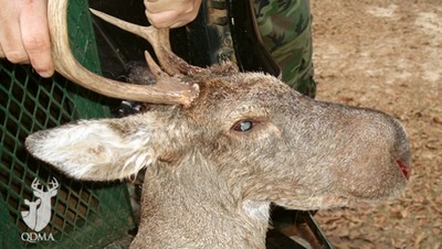
The term “Bullwinkle disease” sounds more like a joke about someone than an actual ailment. As it turns out, Bullwinkle disease in deer is a thing. It’s a true-to-life disease that can impact deer. Although quite rare, it seems white-tailed deer can get an infection that causes their muzzle to swell. In turn, their face looks more like the cartoon moose Bullwinkle than that of a normal, healthy whitetail.
Wildlife vets know that the head swelling is caused by a long-term bacterial infection in soft tissues of the afflicted deer’s face. However, the most fascinating part of Bullwinkle disease is that no one knows how deer get it. Or even where the bacteria comes from.

Bullwinkle Deer
Source: “The Southeastern Cooperative Wildlife Disease Study (SCWDS) has been studying the parasites and diseases of white-tailed deer for more than 56 years. With so much time and effort invested in this area, one would think that few surprises would be left, but that doesn’t ever seem to be the case. Since 2005, we have received samples from ten deer with oddly deformed muzzles, as well as reports of several other affected deer. The swollen muzzles give them a strange appearance and prompted someone to call them “Bullwinkle deer,” based on their resemblance to the 1960’s cartoon character.
Although the cases reported to us are uncommon, they occur over a wide geographic area. In fact, affected white-tailed deer have been submitted to SCWDS from as far north as Michigan and as far south as Alabama. Furthermore, the condition also has been confirmed in a mule deer buck in Idaho.
Bullwinkle Disease in Deer & Head Swelling
The swollen faces are the result of chronic inflammation in the soft tissues of the muzzle. Deer with lumpy jaw can also have swollen jaws, but not to the same extent. The inflammation also is seen in connective tissues in the oral cavity, but it is much more severe on the nose and upper lip. All of the deer examined have had similar colonies of bacteria within the inflammatory infiltrates. Attempts to culture the bacteria have been frustrating. This is possibly due to chronicity of lesions, freezing and storage of samples prior to submission. Alternatively, it may be due to excessive growth of secondary bacterial contaminants.
Staining characteristics and DNA sequencing of the bacterial colonies observed suggest they differ from other organisms known to cause problems in deer. Investigations continue into the bacteria’s potential role in the development of this condition.
So far, all of the reported cases have been in hunter-killed deer or deer observed in the wild. Some deer have been thin, but there have been no reports of mortality directly attributed to this disease. One landowner reported having seen the same affected deer at a backyard feeder for nearly two years. Many of the deer observed or killed have been known to visit feed sites. However, the association with feeding is anecdotal. At this time, we do not know the factors that may predispose a deer to develop this unusual condition.
The lesions are certainly dramatic, but this disease does not appear to have any negative consequences for deer populations. Cases are relatively infrequent and are not clustered. It is possible that this problem has always occurred in deer, but at a very low prevalence. However, it has become very easy for photographs to be widely circulated among hunters and biologists in a very short period of time. We can attribute that to hunters, trail cameras and the internet.
This rapid sharing of information may have increased the detection and submission of rare and unusual cases, such as the Bullwinkle disease in deer we see here. Prepared by Kevin Keel, University of California-Davis”


